EDITOR BOARD MEMBERS :  VIEW ALL

Dr Bikasih Thapa & Dr Maheswar Prasad (Nepal) - Hideo Wada MD PhD (japan) - Dr a Lavra Castrocatesana (Mexico) - Dr Mrs N.M. Hettiarachechui (Srilanka) - Dr Jorge Aldrete Velasco (Mexico) - Prof Hans Peter Kohler (Switzerland) - Dr Hermanus Suhartono S Sp.OG(K) PhD - Dr Isabel Pinheiro (Portugal) - Dr Suranga (Srilanka) - Jovia Dino Jansen Amsterdam,Holand - Hideo Wada MD PhD University Graduate School of Medicine Departement of Moleculer and Laboratory Medicine Japan - DR Bikash Thapa Internal Medicine Nepal University - DR Maheswar Prasad Internal Medicine Nepal University - Dr a Lavra Castro Castresana Colegio de Medicina interna de Mexico - Dr Suransa Manilgama University of Srilanka Internal Departement Medicine - Dr Mrs N.M. Hettiarachechui University of Medicine Srilanka - Dr Jorge Aldrete Velaso .Colegio de Medicina Interna de Mexico - Prof Hans Peter Kholer M.D FACD Profesor of Medicine University ot Switzerland - Dr Ramezan Ali Atace . Baqiyatallah University of Medical Sciences Departement of Micrology Tehran Iran - Ezekiel Wong Toh Yoon Dr. Gastroenterology of Japan - D Eric Beck,MD Bethesda Hospital Capitol Boelevard St Paul USA - Dr Emine Guderen Sahin Istambul University of Internal Medicine Turky - Dr Selmin Toplan Istambul University - Dr Nicholas New Australia - Dr Kughan Govinden. Tropical Infection of Internal Medicine Malaysia - Dr Godfrey M Rwegerera Princes Marina Hospital Bostwana -

Title : MRI PICTURE OF A HUMAN BRAIN FOR AREA DETECTION FOR BRAIN TUMOR

Author : VARADA DEEPTHI, K BALAJI SUNIL CHANDRA, JANGILI RAVI KISHORE

Abstract :

It takes a lot of time and effort to identify, segment, and detect the affected region in a brain tumour. Magnetic resonance imaging (MRI) is a notion in image processing that allows one to see the various human body structures. Using standard imaging methods to detect aberrant brain regions is a huge challenge. Multiple imaging modalities are used in an MRI procedure to examine and record the brain's interior structure. This article focuses on a method for removing noise from medical photographs, and then how to improve them using a balance contrast enhancement technique (BCET) so that an accurate diagnosis may be made. The next step is to apply picture segmentation. The last step is to use the cunning edge detection approach to pick up on the subtle contours. To prove that the suggested method works, the results of the experiment showed that the area of the tumour and normal brain areas could be detected with approximately 98% accuracy in MRI pictures.

[ PDF ]

Editor Board
Editor in chief

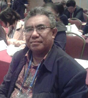
Dr. Arend L Mapanawang, Sp.PD, FINASIM, PhD

Subject Area

Every article submitted to IJHMCR is screened by Turnitin software.

Indexing

PENGUNJUNG KAMI DARI BERBAGAI BELAHAN DUNIA